Cancer-Associated Retinopathy With Thymic Carcinoma
A multimodal approach to treatment led to meaningful visual improvement despite poor prognostic signs on OCT.
Mena Kozman, MD; Aaron Shi, MD; Nyaari Kothiya; Sami Dahr, MD; and Rabia Karani, MD, MPH
Retina Today 
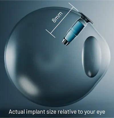
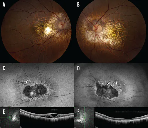
KEY TAKEAWAYS Cancer-associated retinopathy (CAR) is a rare paraneoplastic autoimmune retinopathy characterized by progressive vision loss, for which there is no standard treatment approach. The authors describe a case of CAR in a patient with thymic carcinoma who was treated with an aggressive, multimodal approach including high-dose systemic corticosteroids, intravenous immunoglobulin, sub-Tenon corticosteroids, plasmapheresis, rituximab, intravitreal dexamethasone implant (Ozurdex, Abbvie), and systemic chemotherapy. Despite a poor prognosis, the patient experienced an improvement in functional vision, highlighting both a potential dissociation between structural damage and functional capacity and the importance of aggressive treatment. Cancer-associated retinopathy (CAR) is a rare paraneoplastic autoimmune retinopathy characterized by progressive vision loss. It develops from the cross-reactivity of tumor antigens and retinal proteins, leading to photoreceptor and bipolar cell damage.1-3 First described in association with small-cell lung cancer, CAR has since been reported with gynecologic, hematologic, breast, prostate, and hepatocellular cancers.4 The diagnosis remains challenging, however, as many patients exhibit minimal fundoscopic abnormalities; thus, the combination of imaging and antibody testing can aid in accurate diagnosis. There is no standardized treatment strategy for CAR due to its rarity and heterogeneous presentation. Here, we present a case of CAR in thymic carcinoma treated with a multimodal approach, highlighting the value of aggressive intervention despite profound vision loss. CASE REPORT A 54-year-old man presented to an outside facility with rapidly progressive, painless, bilateral vision loss over 1 week. His VA was hand motion OU and IOP was 8 mm Hg OU with no relative afferent pupillary defect. The anterior segment examination was unremarkable, while the fundus examination revealed attenuated, sclerotic ghost vessels and temporal optic disc pallor in each eye.