Pearls for Managing Complex Diabetic Vitrectomy
Diabetic tractional retinal detachments are challenging for even the most seasoned surgeons. Following these steps can help minimize complications.
Neil Sheth, MD, MBA, and Dimitra Skondra, MD, PhD
Retina Today 
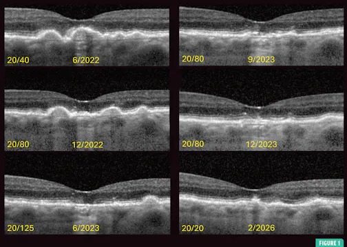
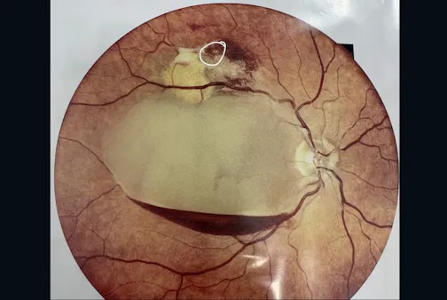
Key Takeaways When planning surgery for tractional retina detachments (TRDs), OCT imaging, ultrasound (when needed), possible cataract extraction, and anti-VEGF injection can help map out the extent of the traction, assess the status of the hyaloid, and regress active neovascularization. Useful tools in TRD surgery include a delaminating spatula, a high-magnification contact lens, MPC vertical scissors, a lighted pick, a Finesse Flex Loop (Alcon), and dextrose added into the infusion bottle. Tamponade selection should favor gas over silicone oil in most complex traction/rhegmatogenous RD cases, as long-acting gas provides 6 to 8 weeks of tamponade and avoids the need for oil removal surgery. Diabetic tractional retinal detachments (TRDs) are some of the most technically demanding cases in vitreoretinal surgery. Each eye presents a unique constellation of posterior hyaloid configuration, vitreoretinal adhesions, fibrovascular proliferation, and neovascular activity. Success demands technical proficiency, systematic preoperative planning, thoughtful selection of instrumentation, and sound intraoperative decision making. In this article, we share our considerations during the key phases of complex diabetic vitrectomy (Table).1 PREOPERATIVE PLANNING Preoperative OCT is invaluable for mapping the extent of the traction, evaluating the foveal contour (including the presence of an epiretinal membrane), identifying focal fibrovascular pegs overlying the macula (and an associated plane for dissection), and identifying any associated tractional-rhegmatogenous component. The integrity of the external limiting membrane and ellipsoid zone can also inform counseling on visual prognosis, which is often limited. When media clarity is compromised by dense vitreous hemorrhage, B-scan ultrasonography is essential. Key features to assess using this tool include the configuration of the posterior hyaloid, the extent of the subhyaloid hemorrhage, the presence and location of a funnel detachment, and the relationship of fibrovascular tissue to the disc.