


Key Takeaways
- Nidek has launched the RSD-1 Duo OCT/Fundus Camera, AFC-1 Non-mydriatic Auto Fundus Camera,and EyeLink Eye Care Data Management Platform
- The RSD-1 Duo combines expanded OCT imaging and fundus photography with PC-free analysis, reference database capabilities and optional OCT angiography
- EyeLink centralizes imaging data and, with the optional Retina Viewer, provides advanced OCT and OCT angiography analysis, including deep learning-based segmentation and longitudinal image comparison
Nidek announced the launch of 3 ophthalmic technologies designed to streamline imaging, screening, analysis and data management: the RSD-1 Duo OCT/Fundus Camera, AFC-1 Non-mydriatic Auto Fundus Camera, and EyeLink Eye Care Data Management Platform.
The company plans to showcase the new products at the European Society of Cataract and Refractive Surgeons (ESCRS) 2026 meeting, scheduled for September 11-14 in London.
The RSD-1 Duo combines optical coherence tomography (OCT) and fundus photography, while the AFC-1 provides nonmydriatic fundus imaging. According to Nidek, both systems incorporate automated operation intended to support consistent image acquisition while reducing operator workload.
For color fundus imaging, the RSD-1 Duo and AFC-1 use slit-scan technology designed to produce high-contrast, high-definition images while reducing artifacts and shadows. The systems provide 50° imaging in pupils as small as 2.0 mm and can automatically combine as many as 7 images to create a panoramic composite. Fundus autofluorescence imaging is also available with the FAF model of the RSD-1 Duo.
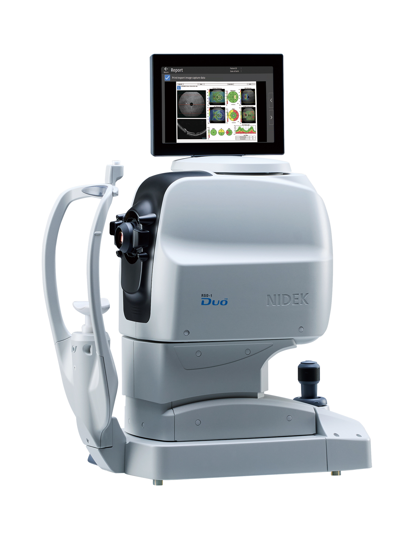
The RSD-1 Duo expands OCT B-scan imaging to as much as 16.5 mm in width and 3.0 mm in depth. According to the company, the larger imaging area can capture anatomy extending from the optic nerve head to the temporal vascular arcade and accommodate the oblate retinal shape associated with myopic eyes.
The system also incorporates B-scan denoising technology, and optional OCT angiography uses an imaging algorithm designed to suppress artifacts and improve visualization of vascular detail.
Nidek said the RSD-1 Duo can perform basic analysis and generate reports without requiring an external PC. Its retina map captures the optic disc and macula and supports reference database analysis of both the retinal nerve fiber layer and ganglion cell complex from a single OCT image.
For glaucoma assessment in eyes with axial myopia, a long axial length reference database provides analysis incorporating axial-length compensation. The device automatically selects between the regular and long axial length reference databases using an axial-length value derived from OCT image acquisition. The company noted that this calculated value differs from an actual measured axial length.
The standard reference database was developed using data from a sample of Caucasian and Asian patients, whereas the long axial length database was developed from a sample of Asian patients.
Both the RSD-1 Duo and AFC-1 offer PC-free operation and connectivity options that include direct DICOM connectivity and integration with Nidek's new EyeLink platform.
EyeLink is designed to centralize patient imaging data on a server, allowing images acquired with the RSD-1 Duo and AFC-1 to be automatically transferred to the platform and accessed from multiple PCs throughout a clinical practice. An optional Retina Viewer application extends the platform's OCT and OCT angiography analysis capabilities. Its deep learning-based segmentation identifies retinal layer boundaries on B-scan images, which serves as the basis for Nidek's Structural Normality Map. The map is designed to identify and highlight structural abnormalities, including subtle retinal changes, to assist clinicians with image interpretation.
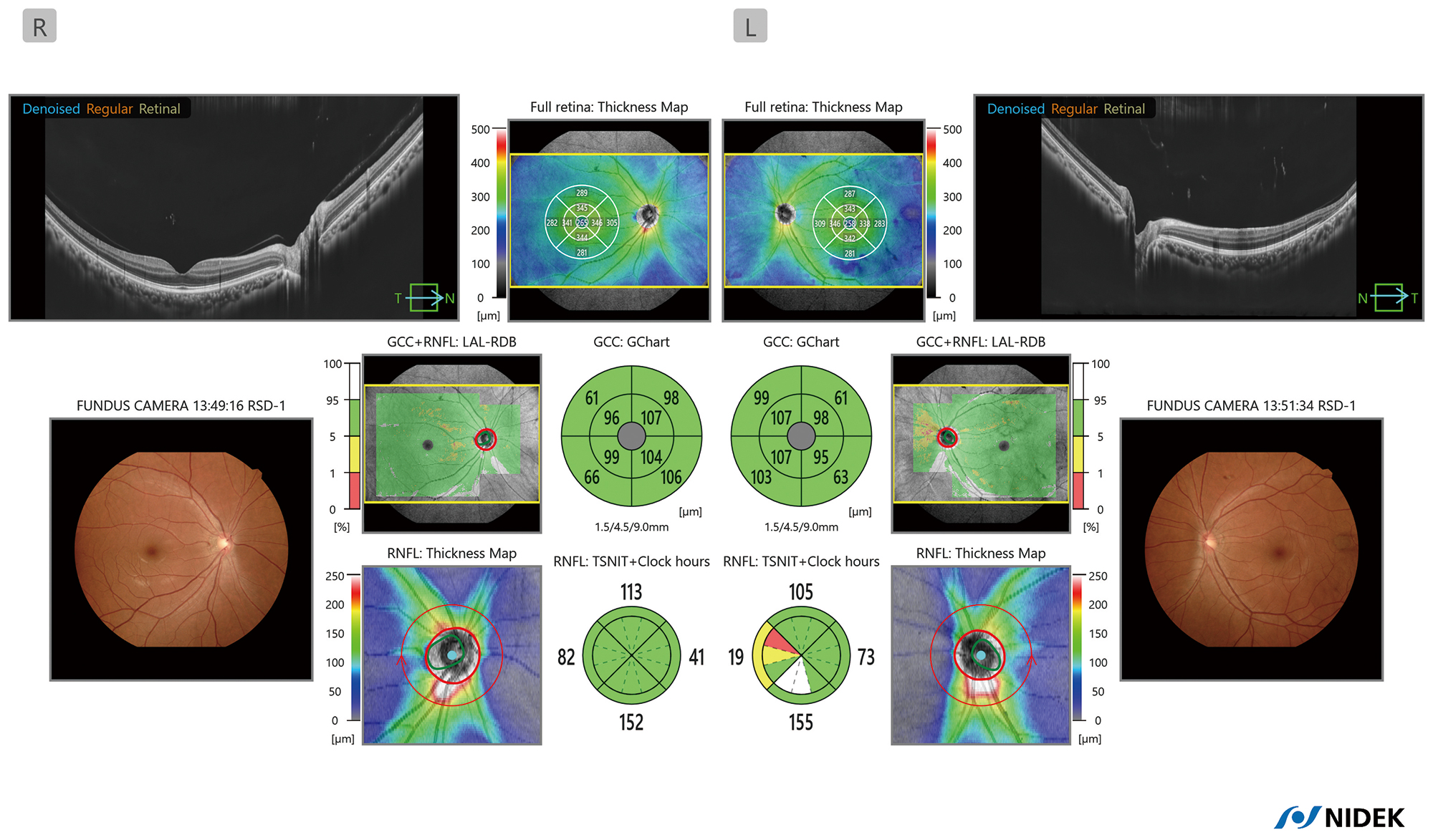
Optional denoising licenses are available for B-scan and OCT angiography images. The viewer also provides quantitative OCT angiography metrics intended to assist with assessment of disease progression and treatment response. For longitudinal evaluation, automatic image registration enables clinicians to compare changes at the same retinal location across examinations, including studies performed with different scan sizes or orientations. Additional tools include anterior chamber angle analysis, pachymetry mapping and epithelial mapping for anterior segment assessment.
"We believe that the RSD-1 Duo and AFC-1, featuring effortless image capture, high-quality imaging, PC-free operation and analysis, and broad connectivity, streamline clinical workflows and support faster, more confident decision-making," Motoki Ozawa, president and CEO of Nidek, said in the company's announcement.
Mr. Ozawa added that EyeLink provides clinicians with a dedicated viewer and advanced analysis capabilities, with Nidek planning to continue expanding the platform.
The company said it will demonstrate the RSD-1 Duo, AFC-1 and EyeLink at booth D.300 during ESCRS 2026.

