


Key Takeaways
- Analysis of retinal imaging from more than 90,000 individuals found that atrial fibrillation was consistently associated with thinning of the macular ganglion cell-inner plexiform layer
- Retinal thinning was detectable an average of 4 years before clinical presentation among individuals who subsequently developed atrial fibrillation
- Researchers said retinal imaging could eventually complement, rather than replace, ECG-based diagnosis by identifying patients who may benefit from additional cardiac assessment
Retinal imaging may reveal structural biomarkers associated with atrial fibrillation (AF) as early as 4 years before the condition is clinically detected, according to a large-scale study led by researchers at Moorfields Eye Hospital NHS Foundation Trust and the UCL Institute of Ophthalmology and published in PLOS Digital Health.
The investigators analyzed retinal imaging data from more than 90,000 individuals across 2 datasets and identified consistent differences in retinal structure among people with AF compared with those without the heart rhythm disorder. The findings suggest that routinely acquired ophthalmic images could eventually help identify patients who may benefit from further cardiovascular assessment.
AF is the most common heart rhythm disorder and affects more than 1.5 million people in the United Kingdom. The condition substantially increases the risk of stroke, heart failure, and death. Because episodes of AF can be intermittent, however, the arrhythmia may not be captured during a standard electrocardiogram (ECG), allowing some cases to remain undiagnosed until complications develop.
The researchers sought to determine whether retinal imaging already routinely performed in community optometry practices and hospital eye clinics could provide a noninvasive means of identifying individuals at increased risk for AF.
Retinal Thinning Associated with AF
The investigators examined optical coherence tomography (OCT) scans and color retinal photographs from the AlzEye and UK Biobank datasets.
The AlzEye dataset included more than 50,000 patients from Moorfields, of whom 5,735 had AF. The UK Biobank cohort included approximately 40,000 participants, including 526 with AF.
Across both datasets, AF was consistently associated with a thinner macular ganglion cell-inner plexiform layer (mGCIPL). Greater thinning of this retinal layer was associated with an increased likelihood of an AF diagnosis.
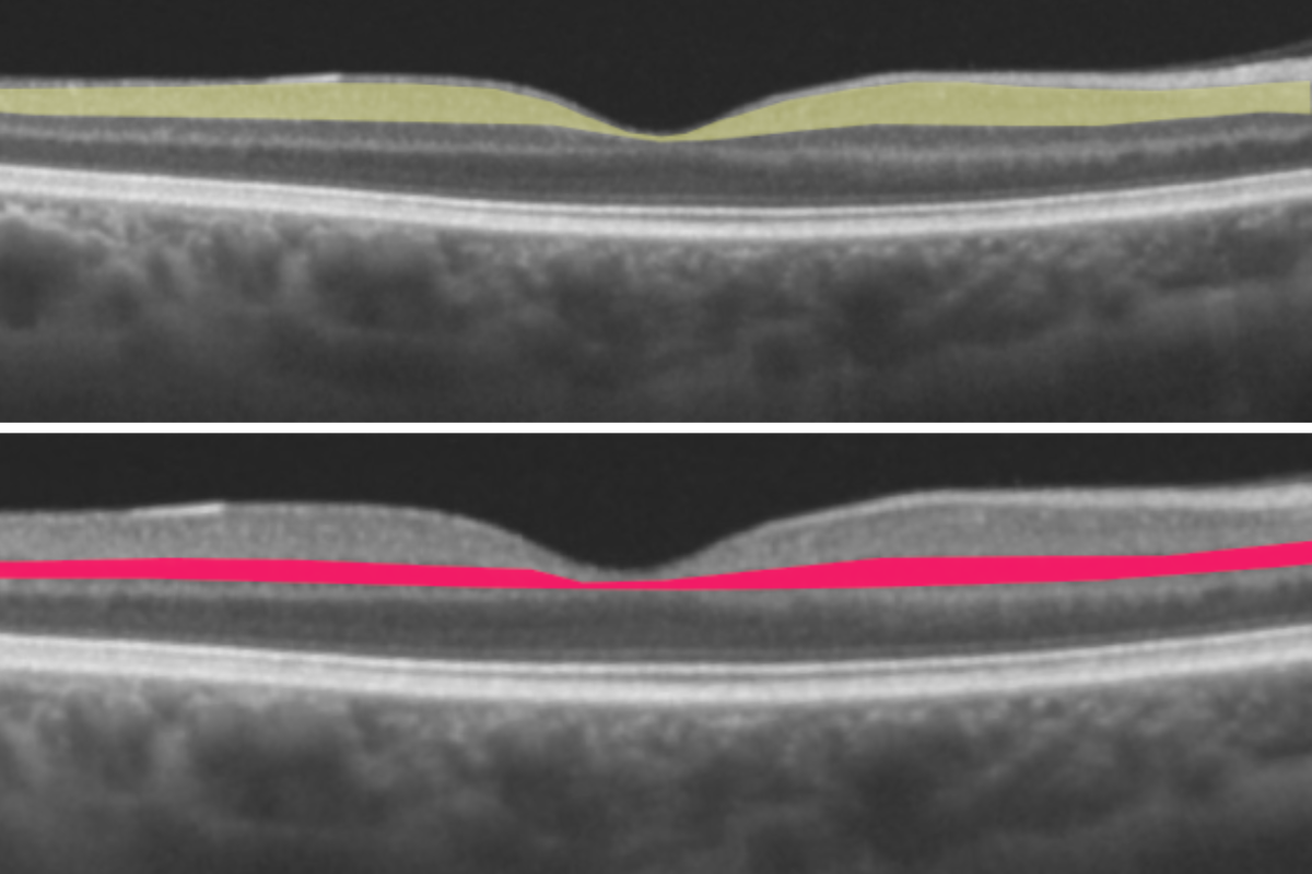
Importantly, the association extended to individuals who did not have AF at the time their retinal images were obtained. Among patients who subsequently developed the condition, the mGCIPL was thinner an average of 4 years before they presented to a hospital with AF. The relationship persisted after the researchers accounted for potential confounding factors including age, sex, ethnicity, hypertension, diabetes, smoking, and alcohol use, according to the study.
The investigators said the results indicate that retinal structural changes associated with AF may precede clinical presentation, potentially providing an opportunity to identify individuals at risk while the disorder remains asymptomatic.
The biological mechanism underlying the association remains uncertain. The study authors proposed that mGCIPL thinning could reflect cerebral microvascular injury, including small strokes occurring as a consequence of undiagnosed AF. Such events could reduce blood flow to retinal nerve cells and contribute to retinal thinning.
“This large-scale analysis shows that retinal imaging can pick up on structural changes within the eye that are clearly linked to AF,” senior author Siegfried Wagner said in a statement. “For patients, the value of this kind of eye imaging is that it is easy to access, quick and doesn't involve any invasive procedures.”
Mr. Wagner emphasized that retinal imaging would not be intended to replace ECG-based diagnosis. Instead, the approach could potentially serve as a complementary screening tool to identify individuals who warrant further cardiac evaluation.
Lead author Josef Huemer noted that additional research will be required before the findings can be translated into clinical screening.
“If confirmed in further research, our findings raise the possibility that routine eye examinations could, in future, help identify people who would benefit from closer cardiac assessment, potentially enabling earlier treatment with therapies known to reduce the risk of stroke,” Mr. Huemer said.
